Your eyes are fascinating organs, designed to capture light and convert it into images. This complex process begins with the cornea, the transparent outer layer that helps focus light. Behind it lies the iris, which controls the amount of light entering by adjusting the size of the pupil.
Light then passes through the lens, which changes shape to focus on objects at varying distances. This focused light reaches the retina, where millions of photoreceptor cells—rods and cones—convert light into electrical signals. These signals travel through the optic nerve to your brain, creating the images you see.

The retina is especially crucial, as it contains the macula, responsible for central vision and fine detail. Surrounding the retina is the vitreous, a clear gel that maintains the eye’s shape. Understanding these components helps explain how your eyes function as a cohesive system to enable vision.
Basic Anatomy and Structure of Your Eye
Understanding the structure of your eye is key to appreciating how vision works. The eye is made up of both external and internal parts, each playing a vital role in how you see the world.
External Features: Iris, Cornea, Sclera, and Pupil
The external features of your eye are designed to protect and regulate light entry. The cornea, a transparent layer at the front, helps focus light. Behind it is the iris, which controls light entry by adjusting the pupil size. The sclera, the white part, provides structural support and protection to the eye. These external layers work together to ensure clear vision and protect your eye from harm.

Internal Components: Lens, Retina, and Vitreous
Inside your eye, the lens focuses light onto the retina, where images are formed. The vitreous, a clear gel, fills the space between the lens and retina, maintaining the eye’s shape. This intricate system ensures that light is properly focused, allowing you to see clearly.
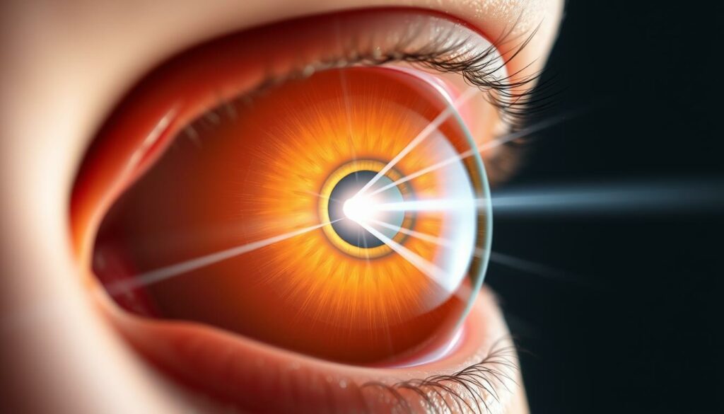
How Light is Focused: From Cornea to Retina
The process of focusing light in your eye is a remarkable journey from the cornea to the retina. This intricate process ensures that light is precisely focused, enabling clear vision.
The Role of the Cornea and Lens
The cornea, the transparent outer layer, is the first to refract incoming light. This refraction is crucial as it bends light before it passes through the lens. The lens then adjusts its shape, controlled by ciliary muscles, to focus light accurately on the retina. This dynamic adjustment ensures sharp images, whether you’re looking at something nearby or far away.
Pupil Regulation and Iris Function
The iris controls the amount of light entering the eye by adjusting the pupil’s size. In bright conditions, the pupil constricts to let in less light, while in low light, it dilates to allow more light in. This regulation is essential for maintaining optimal light levels for clear vision.
Exploring the Different Types of Eyes
Understanding the diversity in eye structures can reveal fascinating insights into how vision has evolved. From simple to complex designs, each type of eye has unique adaptations that enhance its functionality.
Simple Eyes versus Compound Eyes
Simple eyes, like those in humans, use a single lens to focus light onto a retina. This design excels at capturing detailed images, making it ideal for tasks requiring sharp vision. On the other hand, compound eyes, found in insects, consist of thousands of small lenses. These allow for wide-angle vision and rapid motion detection, crucial for navigating dynamic environments.
- Simple eyes focus light through a single lens for detailed vision.
- Compound eyes use multiple lenses for wide field of view and motion detection.
Evolutionary Adaptations in Vision Systems
Evolution has shaped eyes to meet specific needs. The ciliary body adjusts the lens shape, enabling focus on objects at varying distances. Cones in the retina enhance color vision, while rods handle low-light conditions. Over time, these adaptations have optimized vision for different environments.
- The ciliary body and cones improve focus and color perception.
- Surface curvature affects how images project onto the retina.
- Brain processing turns light signals into coherent images.
Vision systems adapt to environments, whether through a wide field of view or detailed central vision. These evolutionary changes highlight nature’s ingenuity in optimizing vision for survival and function.
The Connection Between Your Eyes and Your Brain
Have you ever wondered how the images you see are created? The journey from light entering your eyes to the images formed in your brain is a fascinating process. At the heart of this connection is the optic nerve, a bundle of nerve fibers that carries electrical signals from the retina to the brain.
When light enters your eyes, it’s focused by the cornea and lens onto the retina. The retina’s photoreceptor cells—rods and cones—convert this light into electrical signals. These signals travel through the optic nerve to the brain, where they’re processed to create the images you see.
Optic Nerve and Visual Processing
The optic nerve plays a vital role in vision. It transmits signals from the retina to the brain, where they’re interpreted. The brain processes these signals, allowing you to recognize shapes, colors, and details. This complex process happens quickly, often without you even noticing.
The optic nerve consists of over a million nerve fibers. These fibers carry signals from the retina to the brain. Damage to the optic nerve can lead to vision problems, such as blurred vision or loss of peripheral vision. Conditions like glaucoma can damage the optic nerve, emphasizing the importance of regular eye exams.
Regular eye exams can detect potential issues early. They help protect both your vision and brain health. A healthy optic nerve ensures clear vision and proper communication between your eyes and brain.
Recognizing and Managing Common Eye Conditions
Understanding and addressing common eye conditions is crucial for maintaining healthy vision. Many conditions, such as astigmatism, cataracts, and glaucoma, can affect your ability to see clearly. Recognizing early signs and symptoms is the first step in managing these issues effectively.
Identifying Signs and Symptoms
Common symptoms of eye conditions include blurred vision, irritation, and unusual light sensitivity. For instance, glaucoma often causes peripheral vision loss, while cataracts may lead to cloudy or dim vision. Regular eye tests, such as slit-lamp examinations and tonometry, help diagnose these conditions early. Early detection is key to preventing vision loss and ensuring proper treatment.
Treatment Options: From Medications to Surgery
Treatment depends on the type and severity of the condition. For example, astigmatism can be corrected with eyeglasses or contact lenses, while cataracts may require surgery to remove the cloudy lens. Glaucoma is often managed with eye drops or laser therapy to reduce pressure in the eye. In severe cases, surgery may be necessary to preserve vision.
Regular eye exams are essential for identifying potential problems early. Conditions like diabetic retinopathy and macular degeneration can progress without noticeable symptoms, making routine testing crucial. By understanding your risk factors and staying proactive, you can protect your vision and maintain healthy eyes for years to come.
Effective Strategies for Healthy Eyes
Protecting your vision is a lifelong commitment that requires attention to daily habits and regular check-ups. By adopting simple yet effective care routines and staying proactive about eye exams, you can maintain healthy vision for years to come.
Daily Care Routines and Protective Measures
A consistent daily routine is key to preserving your eye health. Start by practicing good hygiene—wash your hands before touching your eyes and avoid sharing makeup or contact lenses. Ensure you get enough rest, as prolonged screen time can strain your eyes. Follow the 20-20-20 rule: every 20 minutes, look at something 20 feet away for 20 seconds. This can reduce eyestrain, especially if you work on a computer.
Protective eyewear is another crucial aspect. Wear sunglasses with 100% UV protection when outdoors to shield your eyes from harmful UV rays. If you work in environments with dust or chemicals, use safety goggles to prevent injuries. Proper lighting is also important—avoid harsh glares and ensure your workspace is well-lit to reduce strain.
Using Tests and Exams to Monitor Vision
Regular eye exams are essential for detecting potential issues early. The US Preventive Services Task Force recommends that children have at least one eye exam between ages 3 and 5. Adults over 40 should start regular screenings, while those over 60 should have a dilated eye exam every 1 to 2 years. If you have diabetes or a family history of eye diseases, annual exams are advisable.
During a comprehensive eye exam, your eye care professional will check for signs of conditions like glaucoma, cataracts, and age-related macular degeneration. They will assess your visual acuity, measure eye pressure, and examine the retina and optic nerve. Early detection of these issues can prevent vision loss and ensure timely treatment.
| Age Group | Recommended Exam Frequency | Key Tests |
|---|---|---|
| Children (3-5 years) | At least once | Visual acuity test, eye alignment check |
| Adults (40+ years) | Every 1-2 years | Comprehensive dilated exam, glaucoma screening |
| Seniors (60+ years) | Every 1-2 years | Dilated exam, macular degeneration screening |
| Diabetics | Every year | Diabetic retinopathy screening |
By combining daily care routines with regular exams, you can safeguard your vision and maintain healthy eyes for the long term. Remember, proactive care is the best way to protect one of your most valuable senses.
A How-To Guide for Maintaining Optimal Eye Health
Maintaining healthy vision involves more than just visiting an eye doctor when you notice a problem. It requires a proactive approach that includes regular exams, lifestyle adjustments, and preventive care. By understanding how to care for your vision, you can reduce the risk of eye conditions and preserve your ability to see the world clearly.
Scheduling Regular Eye Exams and Refraction Tests
Regular eye exams are essential for detecting potential issues early. These exams include tests that evaluate the health of your cornea, lens, and retina. A refraction test, for example, helps determine the correct prescription for glasses or contact lenses, ensuring your vision remains sharp. Even if you don’t notice any problems, routine check-ups can uncover subtle changes in your eye structure that might indicate future issues.
Adopting Preventative Practices and Lifestyle Changes
Protecting your vision starts with simple, everyday habits. Use proper lighting to avoid straining your eyes, and wear protective eyewear when engaging in activities that could cause injury. Don’t forget to follow the 20-20-20 rule: every 20 minutes, look at something 20 feet away for 20 seconds to reduce eyestrain. Additionally, managing screen time and incorporating rest periods can make a significant difference in long-term eye care.
By combining these strategies, you can safeguard your vision and maintain healthy eyes for years to come. Remember, proactive care is the best way to protect one of your most valuable senses.
Conclusion
Understanding how your eye works is the first step toward effective care and long-term vision preservation. From the intricate anatomy to daily care practices, every element plays a role in maintaining healthy vision. The lens, retina, and optic nerve work together to process light and transmit images to your brain, creating the world you see.
Regular tests and exams are crucial for early detection of potential conditions. Conditions like glaucoma or cataracts can be managed effectively if caught early, preserving your vision. Remember, proactive care is essential for overall health and well-being.
By understanding the connection between your eye structure and daily habits, you can take steps to protect your vision. Simple practices, like following the 20-20-20 rule and wearing protective eyewear, make a significant difference. Stay proactive and prioritize your eye health as part of your wellness routine.
FAQ
How does the retina process light to create vision?
The retina, located at the back of the eye, contains specialized cells called cones and rods. These cells capture light and convert it into signals, which are then transmitted to the optic nerve and eventually to the brain. This process allows you to perceive images and colors.
What role does the cornea play in focusing light?
The cornea, the transparent outer layer of the eye, plays a crucial role in focusing light. It works alongside the lens to refract light onto the retina, ensuring clear vision. Its curvature helps in bending light rays appropriately for accurate image formation.
How does the pupil adjust to different light conditions?
The pupil, controlled by the iris, adjusts its size to regulate the amount of light entering the eye. In bright conditions, the pupil constricts (becomes smaller) to reduce light intake. In low-light conditions, it dilates (becomes larger) to allow more light in, enhancing vision in darker environments.
What is the function of the optic nerve in vision?
The optic nerve acts as a communication pathway between the eye and the brain. It carries electrical signals generated by the retina to the brain, where these signals are interpreted to create the images you see. Damage to the optic nerve can lead to vision problems.
How do the lens and ciliary body work together to focus on objects at different distances?
The lens, controlled by the ciliary body, changes shape to focus on objects at varying distances. When looking at something nearby, the ciliary body tightens, making the lens thicker. For distant objects, the ciliary body relaxes, flattening the lens. This process is known as accommodation.
What are the common tests used to assess eye health?
Common tests include visual acuity exams, refraction tests, and retinal exams. These assessments help determine the sharpness of your vision, the correct lens prescription, and the health of your retina. Regular eye exams are essential for early detection of potential issues.
How can blood vessels in the eye affect vision?
Blood vessels supply oxygen and nutrients to the eye’s tissues. However, conditions like diabetic retinopathy can cause these vessels to leak or grow abnormally, potentially leading to vision problems. Regular check-ups can help identify such issues early.
What are the symptoms of common eye conditions?
Symptoms can include blurred vision, eye pain, double vision, or sudden flashes of light. If you experience any unusual symptoms, it’s important to seek professional advice to rule out serious conditions that may require medical attention.
How often should you schedule eye exams?
The frequency of eye exams depends on your age and health. Adults with no eye problems should have a comprehensive exam every 1-2 years. Children and those with a family history of eye conditions may need more frequent check-ups to monitor their vision and eye health.

Imagine a world where your glasses double as your speakers, offering a seamless hands-free experience. This innovative technology is now a reality, thanks Read more

Are you looking for a way to combine corrective vision with sun protection? Prescription sunglasses offer the perfect blend of Read more

Imagine a pair of glasses that adapts to your surroundings, changing from clear to dark in response to UV light. Transition Read more

Smart glasses have come a long way from being niche gadgets to becoming mainstream wearable devices. Today, they blend fashion Read more